 Gene counts are sourced from ARCHS4, which provides uniform alignment of GEO samples.
You can learn more about ARCHS4 and its pipeline here.
Select conditions below to toggle them from the plot:
| GROUP | CONDITION | SAMPLES |
|---|---|---|
| Macular hole |
GSM5420703 GSM5420704 GSM5420705 GSM5420706 GSM5420707 GSM5420708 GSM5420709
|
|
| Macular pucker |
GSM5420693 GSM5420694 GSM5420695 GSM5420696 GSM5420697 GSM5420698 GSM5420699 GSM5420700 GSM5420701 GSM5420702
|
|
| RNV |
GSM5420686 GSM5420687 GSM5420688 GSM5420689 GSM5420690 GSM5420691 GSM5420692
|
Submission Date: Jul 06, 2021
Summary: Background: Proliferative diabetic retinopathy (PDR) is hallmarked by the formation of retinal neovascularization (RNV) membranes, which can lead to a tractional retinal detachment, the primary reason for severe vision loss in end-stage disease. The aim of this study was to characterize the molecular and cellular features of RNV in order to unravel potential novel drug treatments for PDR. Methods: A total of 42 patients undergoing vitrectomy for PDR, macular pucker or macular hole (control patients) were included in this study. The surgically removed RNV and epiretinal membranes were analyzed by RNA sequencing, single-cell based Imaging Mass Cytometry and conventional immunohistochemistry. Since macrophages were found to be abundant in RNV tissue, vitreal macrophages, also known as hyalocytes, were isolated from the vitreous of patients with PDR by flow cytometry, cultivated and characterized by immunhistochemistry. A bioinformatical drug repurposing approach was applied, in order to identify novel drug options for end-stage diabetic retinopathy disease. Results: The in-depth transcriptional and single-cell protein analysis of diabetic RNV tissue samples revealed an accumulation of endothelial cells, macrophages and myofibroblasts as well as an abundance of secreted ECM proteins such as SPARC, FN1 and several types of collagen in RNV tissue. The immunohistochemical staining of cultivated vitreal hyalocytes from patients with PDR showed that hyalocytes express α-SMA (alpha-smooth muscle actin), a classic myofibroblast marker. According to our drug repurposing analysis, imatinib emerged as a potential drug option for future treatment of PDR. Conclusion: This study delivers the first in-depth transcriptional and single-cell proteomic characterization of RNV tissue samples. Our data suggest an important role of hyalocyte-to-myofibroblast transdifferentiation in the pathogenesis of diabetic vitreoretinal disease and suggests their modulation as a novel possible clinical approach.
GEO Accession ID: GSE179568
PMID: 34795670
Visualize Samples
 Visualizations are precomputed using the Python package scanpy on the top 5000 most variable genes.
Precomputed Differential Gene Expression
 Differential expression signatures are automatically computed using the limma R package.
More options for differential expression are available to compute below.
Signatures:
No precomputed signatures are currently available for this study. You can compute differential gene expression on the fly below:
Select conditions:
Control Condition
Perturbation Condition
Only conditions with at least 1 replicate are available to select
 Differential expression signatures can be computed using DESeq2 or characteristic direction.
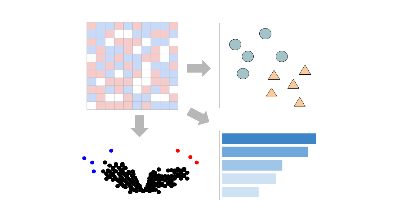
This pipeline enables you to analyze and visualize your bulk RNA sequencing datasets with an array of downstream analysis and visualization tools. The pipeline includes: PCA analysis, Clustergrammer interactive heatmap, library size analysis, differential gene expression analysis, enrichment analysis, and L1000 small molecule search.

 Chatbot
 Single Gene Queries
 Gene Set Queries
 Bulk Studies
 Single Cell Studies
 Hypotheses
 Resources
 Contribute
 Downloads
 About
About
 Help
Help