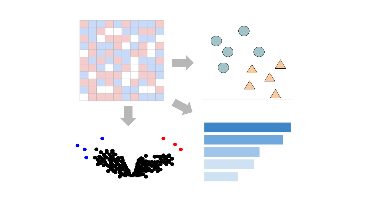
 Gene counts are sourced from ARCHS4, which provides uniform alignment of GEO samples.
You can learn more about ARCHS4 and its pipeline here.
Select conditions below to toggle them from the plot:
| GROUP | CONDITION | SAMPLES |
|---|---|---|
| Skeletal muscle |
GSM3531643 GSM3531644 GSM3531645 GSM3531646 GSM3531647 GSM3531648 GSM3531649 GSM3531650 GSM3531658 GSM3531659 GSM3531660 GSM3531661 GSM3531662 GSM3531663 GSM3531664
|
|
|
GSM3531651 GSM3531652 GSM3531653 GSM3531654 GSM3531655 GSM3531656 GSM3531657 GSM3531665 GSM3531666 GSM3531667 GSM3531668 GSM3531669 GSM3531670 GSM3531671
|
Submission Date: Dec 26, 2018
Summary: Skeletal muscle insulin resistance, decreased phosphatidylinositol 3-kinase (PI3K) activation and altered mitochondrial function are hallmarks of type 2 diabetes. We created mice with a muscle-specific knockout of p110α or p110β, the two major catalytic subunits of PI3K. We find that mice with muscle-specific knockout of p110α, but not p110β, display impaired muscle insulin signaling and reduced muscle size due to enhanced proteasomal and autophagic activity. Despite insulin resistance and muscle atrophy, M-p110αKO mice show decreased serum myostatin, increased mitochondrial mass, increased mitochondrial fusion visualized by intravital microscopy, and increased PGC1α expression, especially PCG1α2 and PCG1α3. This leads to enhanced mitochondrial oxidative capacity, striking increases in muscle NADH content, and higher muscle free radical release measured in vivo using pMitoTimer reporter. Thus, p110α is the dominant catalytic isoform of PI3K in muscle in control of insulin sensitivity and muscle mass, and has a unique role in mitochondrial homeostasis in skeletal muscle.
GEO Accession ID: GSE124394
PMID: 31363081
Visualize Samples
 Visualizations are precomputed using the Python package scanpy on the top 5000 most variable genes.
Precomputed Differential Gene Expression
 Differential expression signatures are automatically computed using the limma R package.
More options for differential expression are available to compute below.
Signatures:
Select conditions:
Control Condition
Perturbation Condition
Only conditions with at least 1 replicate are available to select
 Differential expression signatures can be computed using DESeq2 or characteristic direction.
This pipeline enables you to analyze and visualize your bulk RNA sequencing datasets with an array of downstream analysis and visualization tools. The pipeline includes: PCA analysis, Clustergrammer interactive heatmap, library size analysis, differential gene expression analysis, enrichment analysis, and L1000 small molecule search.

 Chatbot
 Single Gene Queries
 Gene Set Queries
 Bulk Studies
 Single Cell Studies
 Hypotheses
 Resources
 Contribute
 Downloads
 About
About
 Help
Help